4.1.1 Bones, Joints, Action
While I was not present for this activity, my classmates dissected a cow elbow to see how this joint allows movement for the animal. Below is a photo of the dissected cow elbow.
While I was not present for this activity, my classmates dissected a cow elbow to see how this joint allows movement for the animal. Below is a photo of the dissected cow elbow.

4.1.2 Range of Motion
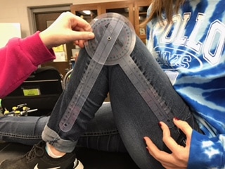
While I was absent for this activity, my classmates were able to measure the range of motion of their joints. They then compared the ranges of motion to analyze what factors have an impact of ROM from individual to individual. Factors include injury, age, weight, activity level, and gender. The student data sheet is also attached below with their measurements and matching joint to picture.
While I was absent for this activity, my classmates were able to measure the range of motion of their joints. They then compared the ranges of motion to analyze what factors have an impact of ROM from individual to individual. Factors include injury, age, weight, activity level, and gender. The student data sheet is also attached below with their measurements and matching joint to picture.

|

|

|
4.2.1 Muscle Rules
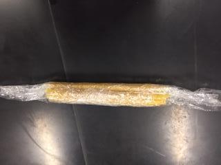
In this activity, our class explored the makeup of a muscle, down to individual muscle fibers. We used spaghetti noodles to illustrate the individual fibers wrapped into a muscle. We then built two muscles on the arm of our Maniken. These muscles were created by following the rules of muscles (Pictured Below).
In this activity, our class explored the makeup of a muscle, down to individual muscle fibers. We used spaghetti noodles to illustrate the individual fibers wrapped into a muscle. We then built two muscles on the arm of our Maniken. These muscles were created by following the rules of muscles (Pictured Below).
|

|

|
4.2.2 Building a Better Body
In this activity, we built the muscles of the chest on our Maniken. These muscles include the intercostal muscles, serratus anterior, pectoralis minor and major, sternal/sternocostalis head, and calvicular head. They can be seen below.
In this activity, we built the muscles of the chest on our Maniken. These muscles include the intercostal muscles, serratus anterior, pectoralis minor and major, sternal/sternocostalis head, and calvicular head. They can be seen below.

4.2.4 Laws of Contraction
While I was not present for this activity, my classmates performed a lab in which they had individual stands of muscle fibers (in test tubes and different solutions) and tested what the effect of ATP on each was. It proved valid that the test tube with the most ATP contracted the least, as ATP allows muscles to relax. Without the presence of ATP, the muscles will contract. The lab can be seen below.
While I was not present for this activity, my classmates performed a lab in which they had individual stands of muscle fibers (in test tubes and different solutions) and tested what the effect of ATP on each was. It proved valid that the test tube with the most ATP contracted the least, as ATP allows muscles to relax. Without the presence of ATP, the muscles will contract. The lab can be seen below.

High magnification picture of a muscle fiber after relaxing from ATP exposure.
|

Low magnification of a muscle fiber that contracted with little to no ATP.
|
4.2.6 You've Got Nerve
In this activity, we created the ulnar and radial nerves of the arm. They are represented by white clay and run from the superior of the Maniken to the fingers. The green clay at the wrist of the Maniken represents the carpal tunnel of the arm, the small space between the wrist and the hand. We also researched the causes and symptoms of carpal tunnel syndrome.
In this activity, we created the ulnar and radial nerves of the arm. They are represented by white clay and run from the superior of the Maniken to the fingers. The green clay at the wrist of the Maniken represents the carpal tunnel of the arm, the small space between the wrist and the hand. We also researched the causes and symptoms of carpal tunnel syndrome.

|

|
4.3.1 The Heart of the Matter
In this activity, we each drew a heart box that shows the blood flow to and from the heart.
Heart Box:
Deoxygenated blood flows from the body to the right atria via the vena cava. From the right atria, the blood flows down the tricuspid valve to the right ventricle, where it is pumped to the lungs via the pulmonary arteries. Here, the blood becomes oxygenated and returns BACK to the heart via the pulmonary veins. The O2 rich blood flows into the left atria to the left ventricle via the bicuspid valve. From the left ventricle, the blood is pumped TO the body via the aorta into arteries of the body.

|

|
4.3.2 Varicose Veins
In this activity, we researched the cause and symptoms of varicose veins. Varicose veins occurs when veins swell and rise to the surface due to increased pressure. More information can be found in the brochure below that I created for this project.
In this activity, we researched the cause and symptoms of varicose veins. Varicose veins occurs when veins swell and rise to the surface due to increased pressure. More information can be found in the brochure below that I created for this project.
| 4.3.2_varicose_veins.pub |
4.3.3 Go With the Flow
In this activity, we created the heart of the Maniken and the vessels that transport blood to and from the heart. The red clay represents oxygenated vessels and the aorta. The blue clay represents deoxygenated blood to the heart from the inferior and superior vena cava.
In this activity, we created the heart of the Maniken and the vessels that transport blood to and from the heart. The red clay represents oxygenated vessels and the aorta. The blue clay represents deoxygenated blood to the heart from the inferior and superior vena cava.

4.3.5 Smoking Can Cause an Arm and a Leg
In this activity, we explored the effects of smoking on the arteries, in relation to PAD (Peripheral Artery Disease) and measured by ABI (Ankle Brachial Index). We chose one partner to have their systolic blood pressure of both their arms and legs be taken and one to record using a blood pressure cuff, stethoscope, and ultrasound machine. We then measured their ABI, seen on the student resource sheet below.
In this activity, we explored the effects of smoking on the arteries, in relation to PAD (Peripheral Artery Disease) and measured by ABI (Ankle Brachial Index). We chose one partner to have their systolic blood pressure of both their arms and legs be taken and one to record using a blood pressure cuff, stethoscope, and ultrasound machine. We then measured their ABI, seen on the student resource sheet below.

While it may be hard to see, her average ABI was 1.12, well above normal.
4.4.1 The Body's Response to Exercise
In this activity, we followed the timeline of a race of a young runner. As the race progressed, we were able to analyze the body systems role in causing her body to fatigue. Her timeline can be seen below.
In this activity, we followed the timeline of a race of a young runner. As the race progressed, we were able to analyze the body systems role in causing her body to fatigue. Her timeline can be seen below.

4.4.2 Mind Over Muscle
While I was not present for this lab, my classmates worked together to measure maximum grip strength and how the muscles of the hand fatigue over time. They used a hand dynometer and EKG sensors connected to Logger Pro System to calculate the grip strength over time. The results of the lab can be seen below.
While I was not present for this lab, my classmates worked together to measure maximum grip strength and how the muscles of the hand fatigue over time. They used a hand dynometer and EKG sensors connected to Logger Pro System to calculate the grip strength over time. The results of the lab can be seen below.

